By Samantha Jones-
Researchers examining brain tissue in both chronic and episodic migraine sufferers have made a major discovery.
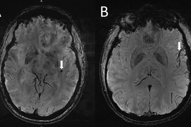
MRI scans show they have enlarged fluid-filled spaces surrounding blood vessels in central regions of the brain and that people who suffer chronic migraines may have problems with the part of the brain responsible for clearing waste.
A perivascular space is a fluid-filled space surrounding certain blood vessels in several organs, including the brain, potentially having an immunological function, but more broadly a dispersive role for neural and blood-derived messengers.
Scientists at the University of Southern California in Los Angeles collected detailed MRI scans from patients suffering from migraines. They found that patients had a higher number of enlarged perivascular spaces, which may be a sign of damage to the small blood vessels of the brain.
Migraines are often preceded or accompanied by other symptoms, such as nausea, fatigue, and a variety of sensory disturbances known as an aura, which can include seeing bright spots of light, ringing in the ears, or numbness and tingling along the body. These episodes typically last for hours, but they can sometimes persist for days to a week.
Migraines are thought to affect about 12% of the population, and women are more likely to report them than men. About 1% to 2% of the population is estimated to experience chronic migraines, or episodes that occur at least 15 days out of the month. They can be acutely managed with pain relievers, and some people have been able to lessen their frequency by avoiding known triggers, like certain foods.
With their new research, USC scientists believe that they’re the first to look at the brains of migraine sufferers using a relatively novel form of ultra-high-resolution MRI known as 7T MRI. They scanned the brains of 20 people with migraines, 10 of whom had chronic migraines and 10 of whom had episodic migraines without aura. For comparison, they also looked at the brains of five healthy controls matched in age.
The study’s authors will be sharing their findings at RSNA 2022 in Chicago, the annual meeting of the Radiological Society of North America (RSNA).
“Perivascular spaces are part of a fluid clearance system in the brain,” study co-author Wilson Xu, an MD candidate at Keck School of Medicine of the University of Southern California in Los Angeles, a neurology researcher said in a statement. “Studying how they contribute to migraine could help us better understand the complexities of how migraines occur.”
‘These changes have never been reported before.’ He added: ‘Perivascular spaces are part of a fluid clearance system in the brain.
Typically, the researchers noted, the fluid-filled spaces are affected by abnormalities at the blood-brain barrier and by inflammation. When they’re enlarged, it can be a sign of underlying small vessel disease.
In the instances of migraine patients, the researchers hypothesized that the enlarged perivascular spaces represent a disruption in the glymphatic system, which is responsible for clearing waste by eliminating soluble proteins and metabolites from the central nervous system.
However, there are still many unknowns surrounding whether the observed changes are a result of migraines, or a contributing cause.
“Although we didn’t find any significant changes in the severity of white matter lesions in patients with and without migraine, these white matter lesions were significantly linked to the presence of enlarged perivascular spaces,” Xu said. “This suggests that changes in perivascular spaces could lead to future development of more white matter legions.”
The study is the first to use 7T MRI to study microvascular changes in the brain due to migraine. There were just 25 participants analyzed: 10 with chronic migraine, 10 with episodic migraine without aura, and five control patients. The authors noted that the small sample size leads to an opportunity for larger studies in the future to learn more.
“The results of our study could inspire future, larger-scale studies to continue investigating how changes in the brain’s microscopic vessels and blood supply contribute to different migraine types,” Xu said. “Eventually, this could help us develop new, personalized ways to diagnose and treat migraine.”

